







X-Ray Diffraction: Comprehensive Guide & Applications (2025)
Introduction of X-ray diffraction
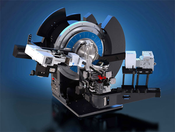
Materials characterization ion is one of the important aspects of the material science field and there are various techniques for analyzing and identifying the material and its composition. One of the techniques which use the principle of X-ray diffraction in characterizing crystalline materials is the X-ray diffraction (XRD) technique. The XRD machine from Bruker is shown in Fig-1.
Fig-1 XRD machine by Bruker [1]
Definition of X-ray diffraction
XRD is a high-precision non-destructive technique that provides important information about the structural characteristics of the materials by enabling chemical composition identification. It provides various information on the crystal structure, phase, crystal orientation, etc of the materials. It works on the principle of diffraction of the X-rays and Its effectiveness depends on the penetration depth of the X-ray beam. This means that diffraction takes place when the light bends slightly as it passes the edge of an object or encounters any obstacles. The degree to which it occurs depends upon the relative size of the wavelength compared to the dimensions of the obstacle or aperture it encounters [2-3]. The schematic representation of the XRD is shown in Fig-2.

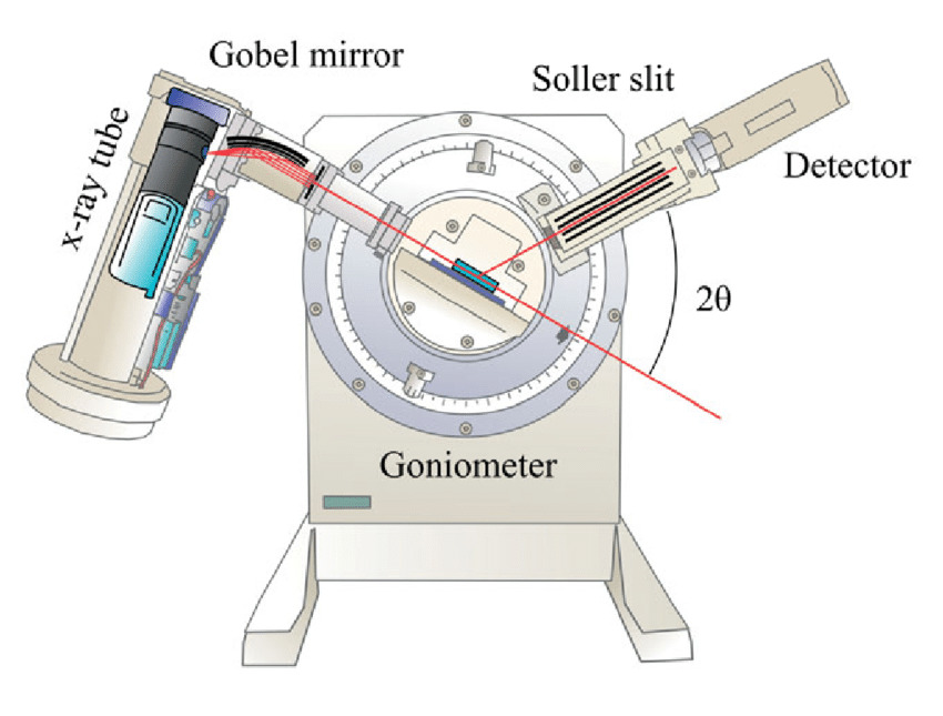
Fig-2 Schematic representation of XRD technique [4]
Working principle of X-ray diffraction
X-rays are a type of electromagnetic radiation, when a monochromatic x-ray scatters from a substance with a structure on this scale, it causes interferences. This interference creates a pattern of lower and higher intensities due to constructive and destructive interference (Bragg’s law). If there is a crystalline substance then a three-dimensional pattern is created like the spacings of planes in the crystal lattice, this process is called constructive interference. By collecting all the diffracted x-rays one can analyze the samples’ structure. The way the X-ray reveals the atomic structure of the crystals is based on Bragg’s law [5]. Fig-3 shows the schematic representation of the XRD working principle.
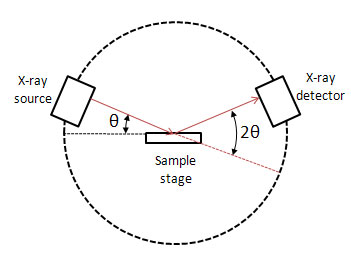

Fig-3 Schematic representation of XRD working principle [6]
Components of X-ray diffraction
The three main components of the XRD are the X-ray tube, sample holder, and X-ray detector.
- X-ray tube: It is the source from which the X-ray is emitted and it consists of a cathode ray tube with a heating tungsten filament. This produces the electrons which accelerate on to the sample surface by applying a voltage. The bombardment of the electrons on the samples is controlled by the filaments which affect the X-ray output intensity.
- Sample holder: it is the basement on which the sample to be characterized is placed and it can be rotated in the direction of the X-ray tube and the detector. The adjustments are made as per the reflection and emission of the X-rays from the sample.
- X-ray detector: The X-ray detector is used to record and process the X-ray signal received from the sample. The signals received are converted into count rate which is obtained as the output on the monitor or printer.
Bragg-Brentano configuration in X-ray diffraction
The Bragg-Brentano configuration is used in most powder diffractometers for parafocusing geometry. It offers very high-resolution and high beam-intensity analysis which requires a very precise alignment. Samples are carefully prepared, and their surface is kept on the tangent plane of the focusing circle, which is defined by three spots at the sample, X-ray source, and receiving slit. incident- and diffracted-beam slits move on a circular plane by centering the sample. Divergent X-rays from the X-ray tube hit the sample at different points on its surface. During the diffraction process, the X-rays are refocused at the detector slit. The schematic representation of the Bragg banteno configuration is shown in Fig-8.
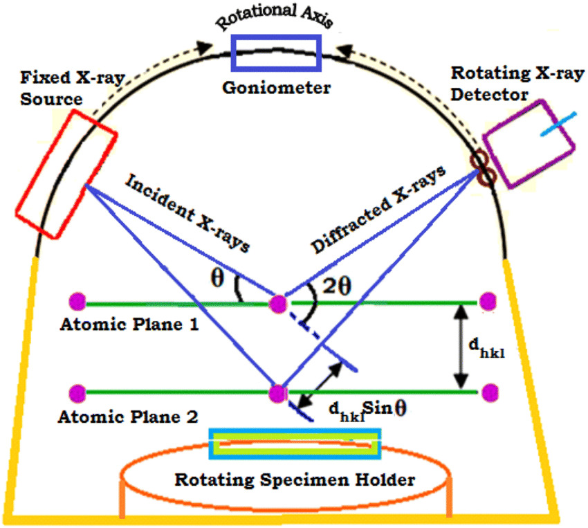

Fig-4 Schematic representation of Bragg banteno configuration [7]
Advantages and Disadvantages of X-ray diffraction
The main advantage of this technique is that it is a nondestructive technique and requires a small sample amount. It helps in identifying the phases and not just the elements, also information about crystallinity, crystal size, and orientation can be obtained. There is no requirement for sample preparation or minimal preparation can be required and does not need vacuum operating conditions.
The major limitations of the XRD are it cannot identify the amorphous materials and does not gives information on profile depth. It is a bulk technique requiring a large analysis volume and a large minimum spot size.
Check this video: https://www.youtube.com/watch?v=JJF_gxdYXEk

Reference
[1] https://blue-scientific.com/bruker-xrd-in-plane-diffraction/
[6] https://xrd.co/component-parts-x-ray-diffractometer/
Nice write up hope this will work perfectly to know the percentage of lithium in an ore